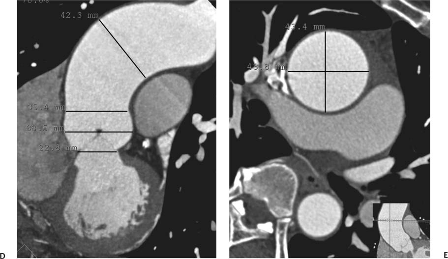


The main limits of CT are related to the X-ray dose exposure and the use of iodinated contrast media. This is why today the employment of a 64 or wider ECG-gated row detector system is suggested.ĬT showing the anomaly of the right subclavian artery (arrow) with an evident retroesophageal course (also called lusory artery) acquisitions require quite short times and nowadays they are almost universally available. Unfortunately, the ECG-gated technique increases the acquisition time as well as the breath-hold required. Thanks to the ECG-gated technique, it is possible to perform the kinetic evaluation of the aortic leaflets and is useful to assess the valve function. Nevertheless, in the thoracic area the ECG-gated technique is strongly suggested to avoid the evidence of false positive flap due to high pulsatility of the root and ascending aorta. Sixteen and wider row detectors provide isotropic pixels, mandatory for the ineludible longitudinal reconstruction. The acquisitions require quite short times and nowadays they are almost universally available. Identification of anatomic variants is also possible ( Figure 1), as well as distinction among acute syndromes. The new generation CTs show sensitivities up to 100% and specificities of 98-99%, allowing the possibility to evaluate the entire aorta including lumen and wall, the possible thrombotic apposition and the peri-aortic area. Fast scanning achieved with the new wide area detector, low artifact sensibility due to fast velocity tube rotation and 24-hour availability in the emergency rooms are the main advantages of CT usage in the medical practice. 2 Computed Tomography (CT) 2.1 Computed Tomography (CT)Ĭomputed Tomography (CT) is currently the most widely employed technique for the study of the thoracic aorta. Nevertheless, differences in spatial resolution, movement artifact susceptibility, acquisition time, contraindications, and risks make a critical re-evaluation of the state of the technology useful to optimize their employment in relation to different clinical situations and apparatus performances. The main limitation of MRI, however, is related to the scarce visibility of stents and calcifications.Īt the present time, both CT and MR imaging are valuable techniques in the study of the thoracic aorta. Nevertheless, since it requires iodinated contrast media and X-ray exposure, it may be adequately replaced by MRI in the follow up of aortic diseases. CT is preferred in the acute aortic disease. The main MRI disadvantages are claustrophobia, presence of ferromagnetic implants, pacemakers, longer acquisition times with respect to CT, inability to use contrast media in cases of renal insufficiency, lower spatial resolution and less availability than CT. Nevertheless, if compared to CT, acquisition times remain longer and movement artifact susceptibility higher. MRI has great potential in the study of the thoracic aorta. The main limits are related to the X-ray dose expoure and the use of iodinated contrast media. The new generation CTs show sensitivities up to 100% and specificities of 98-99%. Nowadays, CT represents the most widely employed technique for the study of the thoracic aorta. At present time, both CT and MRI are valuable techniques in the study of the thoracic aorta.


 0 kommentar(er)
